Among the popular knowledge about early gastric cancer, there are some rare disease knowledge points that require special attention and learning. One of them is HP-negative gastric cancer. The concept of "uninfected epithelial tumors" is now more popular. There will be different opinions on the name issue. This content theory is mainly based on the content related to the magazine "Stomach and Intestine", and the name also uses "HP-negative gastric cancer".
This type of lesions has the characteristics of low incidence, difficulty in identification, complex theoretical knowledge, and the simple MESDA-G process is not applicable. Learning this knowledge requires facing up to the difficulties.
1. Basic knowledge of HP-negative gastric cancer
History
In the past, it was believed that the single culprit in the occurrence and development of gastric cancer was HP infection, so the classic canceration model is HP - atrophy - intestinal metaplasia - low tumor - high tumor - canceration. The classic model has always been widely recognized, accepted and firmly believed. Tumors develop together on the basis of atrophy and under the action of HP, so cancers mostly grow in atrophic intestinal tracts and less normal non-atrophic gastric mucosa.
Later, some doctors discovered that gastric cancer can occur even in the absence of HP infection. Although the incidence rate is very low, it is indeed possible. This type of gastric cancer is called HP-negative gastric cancer.
With the gradual understanding of this type of disease, in-depth systematic observations and summaries have begun, and the names are constantly changing. There was an article in 2012 called "Gastric Cancer after Sterilization", an article in 2014 called "HP-negative Gastric Cancer", and an article in 2020 called "Epithelial Tumors Not Infected with Hp". The name change reflects the deepening and comprehensive understanding.
Gland Types and Growth Patterns
There are two main types of fundic glands and pyloric glands in the stomach:
Fundic glands (oxyntic glands) are distributed in the fundus, body, corners, etc. of the stomach. They are linear single tubular glands. They are composed of mucous cells, chief cells, parietal cells and endocrine cells, each of which performs their own functions. Among them, chief cells The secreted PGI and MUC6 staining were positive, and the parietal cells secreted hydrochloric acid and intrinsic factor;
The pyloric glands are located in the gastric antrum area and are composed of mucus cells and endocrine cells. Mucus cells are MUC6 positive, and endocrine cells include G, D cells and enterochromaffin cells. G cells secrete gastrin, D cells secrete somatostatin, and enterochromaffin cells secrete 5-HT.
Normal gastric mucosal cells and tumor cells secrete a variety of different types of mucus proteins, which are divided into "gastric", "intestinal" and "mixed" mucus proteins. The expression of gastric and intestinal mucins is called a phenotype and not the specific anatomical location of the stomach and intestines.
There are four cell phenotypes of gastric tumors: completely gastric, gastric-dominant mixed, intestinal-dominant mixed, and completely intestinal. Tumors that occur on the basis of intestinal metaplasia are mostly gastrointestinal mixed phenotype tumors. Differentiated cancers mainly show intestinal type (MUC2+), and diffuse cancers mainly show gastric type (MUC5AC+, MUC6+).
Determining Hp negative requires a specific combination of multiple detection methods for comprehensive determination. HP-negative gastric cancer and post-sterilization gastric cancer are two different concepts. For information on the X-ray manifestations of HP-negative gastric cancer, please refer to the relevant section of the "Stomach and Intestine" magazine.
2. Endoscopic manifestations of HP-negative gastric cancer
Endoscopic diagnosis is the focus of HP-negative gastric cancer. It mainly includes fundic gland type gastric cancer, fundic gland mucosal type gastric cancer, gastric adenoma, raspberry foveolar epithelial tumor, signet ring cell carcinoma, etc. This article focuses on the endoscopic manifestations of HP-negative gastric cancer.
1) Fundic gland type gastric cancer
-White raised lesions
fundic gland type gastric cancer

◆Case 1: White, raised lesions
Description: Gastric fundic fornix-greater curvature of the cardia, 10 mm, white, O-lia type (SMT-like), without atrophy or intestinal metaplasia in the background. Arbor-like blood vessels can be seen on the surface (NBI and enlargement slightly)
Diagnosis (combined with pathology): U, O-1la, 9mm, fundic gland type gastric cancer, pT1b/SM2 (600μm), ULO, Ly0, VO, HMO, VMO
-White flat lesions
fundic gland type gastric cancer

◆Case 2: White, flat/depressed lesions
Description: The anterior wall of the gastric fundic fornix-cardia greater curvature, 14 mm, white, type 0-1lc, with no atrophy or intestinal metaplasia in the background, unclear borders, and dendritic blood vessels seen on the surface. (NBI and amplification abbreviated)
Diagnosis (combined with pathology): U, 0-Ilc, 14mm, fundic gland type gastric cancer, pT1b/SM2 (700μm), ULO, Ly0, VO, HMO, VMO
-Red raised lesions
fundic gland type gastric cancer

◆Case 3: Red and raised lesions
Description: The anterior wall of the great curvature of the cardia is 12 mm, obviously red, type 0-1, with no atrophy or intestinal metaplasia in the background, clear borders, and dendritic blood vessels on the surface (NBI and enlargement slightly)
Diagnosis (combined with pathology): U, 0-1, 12mm, fundic gland type gastric cancer, pT1b/SM1 (200μm), ULO, LyO, VO, HMO, VMO
-Red, flat, depressed lesions
fundic gland type gastric cancer
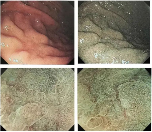
◆Case 4: Red, flat/depressed lesions
Description: Posterior wall of the greater curvature of the upper part of the gastric body, 18mm, light red, O-1Ic type, no atrophy or intestinal metaplasia in the background, unclear boundary, no dendritic blood vessels on the surface, (NBI and enlargement omitted)
Diagnosis (combined with pathology): U, O-1lc, 19mm, fundic gland type gastric cancer, pT1b/SM1 (400μm), ULO, LyO, VO, HMO, VMO
discuss
Males with this disease are older than females, with the average age being 67.7 years old. Due to the characteristics of simultaneity and heterochrony, patients diagnosed with fundic gland type gastric cancer should be reviewed once a year. The most common site is the fundic gland area in the middle and upper part of the stomach (the fundus and the middle and upper part of the gastric body). White SMT-like raised lesions are more common in white light. The main treatment is diagnostic EMR/ESD.
No lymphatic metastasis or vascular invasion has been seen so far. After treatment, it is necessary to determine whether to perform additional surgery and evaluate the relationship between malignant status and HP. Not all fundic gland-type gastric cancers are HP negative.
1) Fundic gland mucosal gastric cancer
Fundic gland mucosal gastric cancer
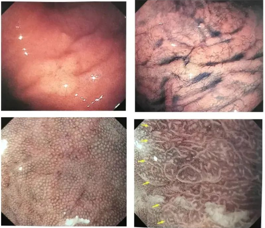
◆Case 1
Description: The lesion is slightly raised, and RAC non-atrophic gastric mucosa can be seen around it. Rapidly changing microstructure and microvessels can be seen in the raised part of ME-NBI, and DL can be seen.
Diagnosis (combined with pathology): Fundic gland mucosal gastric cancer, U zone, 0-1la, 47*32mm, pT1a/SM1 (400μm), ULO, Ly0, VO, HMO, VMO
Fundic gland mucosal gastric cancer
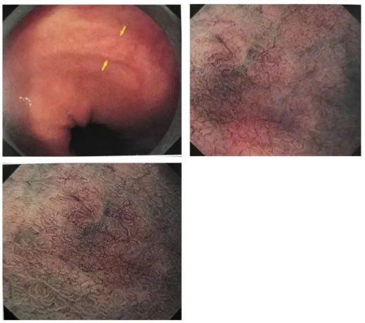
◆Case 2
Description: A flat lesion on the anterior wall of the lesser curvature of the cardia, with mixed discoloration and redness, dendritic blood vessels can be seen on the surface, and the lesion is slightly raised.
Diagnosis (combined with pathology): Fundic gland mucosal gastric cancer, 0-lla, pT1a/M, ULO, LyOV0,HM0,VMO
discuss
The name of "gastric gland mucosal adenocarcinoma" is a bit difficult to pronounce, and the incidence rate is very low. It requires more efforts to recognize and understand it. Fundic gland mucosal adenocarcinoma has the characteristics of high malignancy.
There are four major characteristics of white light endoscopy: ① homochromatic-fading lesions; ② subepithelial tumor SMT; ③ dilated dendritic blood vessels; ④ regional microparticles. ME performance: DL(+)IMVP(+)IMSP(+)MCE widens IP and increases. Using the MESDA-G recommended process, 90% of fundic gland mucosal gastric cancers meet the diagnostic criteria.
3) Gastric adenoma (pyloric gland adenoma PGA)
gastric adenoma
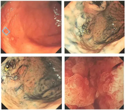
◆Case 1
Description: A white flat raised lesion was seen on the posterior wall of the gastric fornix with unclear boundaries. Indigo carmine staining showed no clear boundaries, and the LST-G-like appearance of the large intestine was seen (enlarged slightly).
Diagnosis (combined with pathology): low atypia carcinoma, O-1la, 47*32mm, well-differentiated tubular adenocarcinoma, pT1a/M, ULO, Ly0, VO, HMO, VMO
gastric adenoma
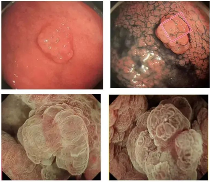
◆Case 2
Description: A raised lesion with nodules on the anterior wall of the middle part of the gastric body. Active gastritis can be seen in the background. Indigo carmine can be seen as the border. (NBI and magnification slightly)
Pathology: MUC5AC expression was seen in the superficial epithelium, and MUC6 expression was seen in the superficial epithelium. The final diagnosis was PGA.
discuss
Gastric adenomas are essentially mucinous glands penetrating the stroma and covered by foveolar epithelium. Due to the proliferation of glandular protrusions, which are hemispherical or nodular, gastric adenomas seen with endoscopic white light are all nodular and protruding. It is necessary to pay attention to the 4 classifications of Jiu Ming under endoscopic examination. ME-NBI can observe the characteristic papillary/villous appearance of PGA. PGA is not absolutely HP negative and non-atrophic, and has a certain risk of canceration. Early diagnosis and early treatment are advocated, and after discovery, active en bloc resection and further detailed study are recommended.
4) (raspberry-like) foveolar epithelial gastric cancer
raspberry foveolar epithelial gastric cancer
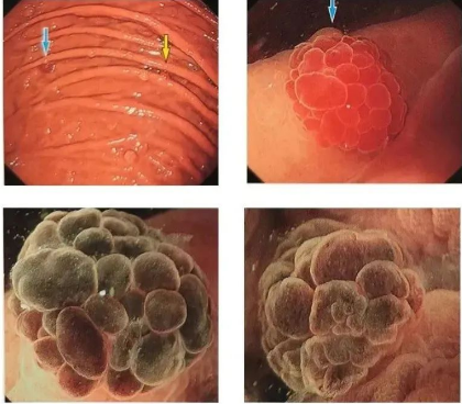
◆Case 2
Description: (omitted)
Diagnosis (combined with pathology): foveolar epithelial gastric cancer
raspberry foveolar epithelial gastric cancer
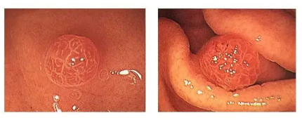
◆Case 3
Description: (omitted)
Diagnosis (combined with pathology): foveolar epithelial gastric cancer
discuss
Raspberry, called "Tuobai'er" in our hometown, is a wild fruit on the roadside when we were children. Glandular epithelium and glands are connected, but they are not the same content. It is necessary to understand the growth and development characteristics of epithelial cells. Raspberry epithelial gastric cancer is very similar to gastric polyps and can easily be mistaken for gastric polyps. The hallmark feature of foveolar epithelium is the dominant expression of MUC5AC. So foveolar epithelial carcinoma is the general term for this type. It can exist in HP negative, positive, or after sterilization. Endoscopic appearance: round bright red strawberry-like bulge, generally with clear borders.
5) Signet ring cell carcinoma
Signet ring cell carcinoma: white light appearance
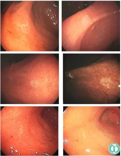
Signet ring cell carcinoma: white light appearance
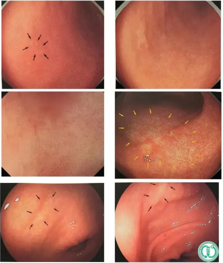
signet ring cell carcinoma
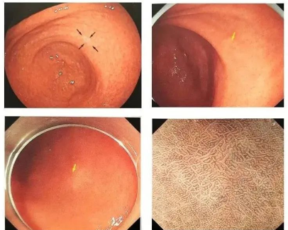
◆Case 1
Description: Flat lesion on the posterior wall of the gastric vestibule, 10 mm, faded, type O-1Ib, no atrophy in the background, visible border at first, not obvious border on reexamination, ME-NBI: only the interfoveal part becomes white, IMVP(-)IMSP (-)
Diagnosis (combined with pathology): ESD specimens are used to diagnose signet ring cell carcinoma.
Pathological manifestations
Signet ring cell carcinoma is the most malignant type. According to the Lauren classification, gastric signet ring cell carcinoma is classified as a diffuse type of carcinoma and is a type of undifferentiated carcinoma. It commonly occurs in the body of the stomach, and is more common in flat and sunken lesions with discolored tones. Raised lesions are relatively rare and can also manifest as erosion or ulcers. It is difficult to detect during endoscopic examination in the early stages. Treatment can be curative resection such as endoscopic ESD, with strict postoperative follow-up and evaluation of whether to perform additional surgery. Non-curative resection must require additional surgery, and the surgical method is decided by the surgeon.
The above text theory and pictures come from "Stomach and Intestine"
In addition, attention should also be paid to esophagogastric junction cancer, cardia cancer, and well-differentiated adenocarcinoma found in HP-negative background.
3. Summary
Today I learned the relevant knowledge and endoscopic manifestations of HP-negative gastric cancer. It mainly includes: fundic gland type gastric cancer, fundic gland mucosal type gastric cancer, gastric adenoma, (raspberry-like) foveolar epithelial tumor and signet ring cell carcinoma.
The clinical incidence of HP-negative gastric cancer is low, it is difficult to judge, and it is easy to miss diagnosis. What is even more difficult is the endoscopic manifestations of complex and rare diseases. It should also be understood from an endoscopic perspective, especially the theoretical knowledge behind it.
If you look at the gastric polyps, erosions, and red and white areas, you should consider the possibility of Hp-negative gastric cancer. The judgment of HP negative must comply with the standards, and attention should be paid to false negatives caused by over-reliance on breath test results. Experienced endoscopists trust their own eyes more. Facing the detailed theory behind HP-negative gastric cancer, we must continue to learn, understand and practice to master it.
We, Jiangxi Zhuoruihua Medical Instrument Co.,Ltd., is a manufacturer in China specializing in the endoscopic consumables, such as biopsy forceps, hemoclip, polyp snare, sclerotherapy needle, spray catheter, cytology brushes, guidewire, stone retrieval basket, nasal biliary drainage catheter etc. which are widely used in EMR, ESD, ERCP. Our products are CE certified, and our plants are ISO certified. Our goods have been exported to Europe, North America, Middle East and part of Asia, and widely obtains the customer of the recognition and praise!
Post time: Jul-12-2024


