

Small ureteral stones can be treated conservatively or extracorporeal shock wave lithotripsy, but large-diameter stones, especially obstructive stones, require early surgical intervention.
Due to the special location of upper ureteral stones, they may not be accessible with a rigid ureteroscope, and stones can easily move up into the renal pelvis during lithotripsy. Percutaneous nephrolithotomy increases the risk of renal bleeding when establishing a channel.
The rise of flexible ureteroscopy has effectively solved the above problems. It enters the ureter and renal pelvis through the normal orifice of the human body. It is safe, effective, minimally invasive, has less bleeding, less pain for the patient, and a high stone-free rate. It has now become a commonly used surgical method to treat upper ureteral stones.
The emergence of the ureteral access sheath has greatly reduced the difficulty of flexible ureteroscopic lithotripsy. However, with the increase in the number of treatment cases, its complications have gradually attracted attention. Complications such as ureteral perforation and ureteral stricture are common. The following are the three major factors leading to ureteral stricture and perforation.
1. Course of disease, stone diameter, stone impaction
Patients with a longer course of disease tend to have larger stones, and large stones remain in the ureter for a long time to form incarceration. Stones at the impaction site compress the ureteral mucosa, resulting in insufficient local blood supply, mucosal ischemia, inflammation and scar formation, which are closely related to the formation of ureteral stricture.
2. Ureteral injury
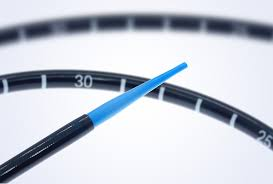
The flexible ureteroscope is easy to bend, and a ureteral access sheath needs to be inserted before lithotripsy. The insertion of the channel sheath is not performed under direct vision, so it is inevitable that the ureteral mucosa will be damaged or perforated due to the bending of the ureter or the narrow lumen during the insertion of the sheath.
In addition, in order to support the ureter and drain the perfusion fluid to reduce the pressure on the renal pelvis, a channel sheath through F12/14 is usually selected, which may cause the channel sheath to directly compress the ureteral wall. If the surgeon's technique is immature and the operation time is prolonged, the compression time of the channel sheath on the ureteral wall will be increased to a certain extent, and the risk of ischemic damage to the ureteral wall will be greater.
3. Holmium laser damage
The stone fragmentation of holmium laser mainly relies on its photothermal effect, which causes the stone to directly absorb the laser energy and increase the local temperature to achieve the purpose of stone fragmentation. Although the thermal radiation depth during the gravel crushing process is only 0.5-1.0 mm, the overlapping effect caused by continuous gravel crushing is inestimable.
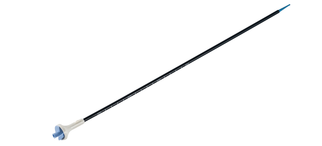
The key points for inserting the ureteral access sheath are as follows:
1. There is an obvious sense of breakthrough when inserting into the ureter, and it feels smooth when it goes up in the ureter. If the insertion is difficult, you can swing the guide wire back and forth to observe whether the guide wire goes in and out smoothly, so as to determine whether the channel sheath is advancing in the direction of the guide wire, such as If there is obvious resistance, the direction of sheathing needs to be adjusted;
The successfully placed channel sheath is relatively fixed and will not come in and out at will. If the channel sheath pops out obviously, it means that it is coiled in the bladder and the guide wire has prolapsed from the ureter and needs to be re-placed;
3. Ureteral channel sheaths have different specifications. Male patients generally use the 45 cm long model, and female or shorter male patients use the 35 cm long model. If the channel sheath is inserted, it can only pass through the ureteral opening or cannot go up to a higher level. Position, male patients can also use a 35 cm introducing sheath, or switch to a 14F or even thinner fascial expansion sheath to prevent the flexible ureteroscope from being unable to ascend to the renal pelvis;
Do not place the channel sheath in one step. Leave 10 cm outside the urethral orifice to prevent damage to the ureteral mucosa or renal parenchyma at the UPJ. After inserting the flexible scope, the channel sheath position can be adjusted again under direct vision.
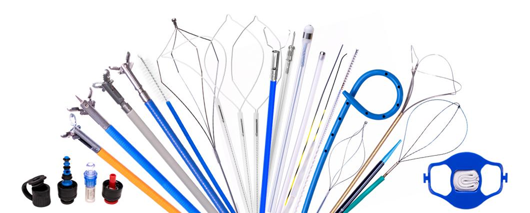
We, Jiangxi Zhuoruihua Medical Instrument Co.,Ltd., is a manufacturer in China specializing in the endoscopic consumables, such as biopsy forceps, hemoclip, polyp snare, sclerotherapy needle, spray catheter, cytology brushes, guidewire, stone retrieval basket, nasal biliary drainage catheter etc. which are widely used in EMR, ESD, ERCP. And Urology Series, such as Nitinol Stone Extractor, Urological Biopsy Forceps, and Ureteral Access Sheath and Urology Guidewire. Our products are CE certified, and our plants are ISO certified. Our goods have been exported to Europe, North America, Middle East and part of Asia, and widely obtains the customer of the recognition and praise!
Post time: Sep-11-2024