
(1). Basic techniques The basic techniques of EMR are as follows:
Sequence of techniques
①Inject the local injection solution just below the lesion.
②Place the snare around the lesion.
③The snare is tightened to grasp and strangulate the lesion.
④Continue to tighten the snare while applying electricity to cut off the lesion.
⑤Retrieve the resected specimen.
(2). Tips
1.Tips for body position selection and endoscope positioning
Because the lesion needs to be treated while the whole image is visible, the patient’s position is very important. Try to twist the scope so that the lesion is located near the biopsy forceps opening, that is, from 5 to 7 o’clock on the screen.
Before treatment, the residue and excess pigment need to be washed and then removed by suction.
For example, if a lesion in the proximal sigmoid colon is removed in the supine or left lateral decubitus position, the specimen will often move to the descending colon, making it difficult to retrieve , so right lateral decubitus position is better for resection.
Likewise, from the perspective of specimen recovery, the left lateral decubitus position is preferred for resection of transverse colon lesions.
2.Tips for local injections
A thick local injection needle can be injected at a lower pressure, but it is not sharp enough and the needle hole is too large, so the author uses a 25G local injection needle.
It is no exaggeration to say that the success or failure of EMR depends largely on local injections.
For small lesions, puncture is performed from the anal side of the lesion to just below the lesion.
For lesions in the curved part or across the folds, if local injection is performed from the anal side, in most cases the lesions become unclear because they are facing the oral side, so local injection should be started from the oral side.
Essentials for Endoscopy Technicians
If liquid leaks out, or there is great resistance during injection, or there is no resistance when liquid enters but no bulge is formed, the injection must be stopped and the operator must be informed of the situation in a timely manner to discuss countermeasures.
The more injection volume, the better.
The trick is to continue injecting as much as possible through a single puncture until the entire lesion is lifted.
3.Tips for choosing a snare
If the snare is in the shape of an elongated oval, the normal mucosa on the lesion mouth and anus sides may be easily and unnecessarily included .
The snare is preferably nearly circular, easy to open laterally, not easy to slide, and has a certain hardness to press on the lesion to grasp the lesion.
The size of the snare should be adapted to the size of the lesion.
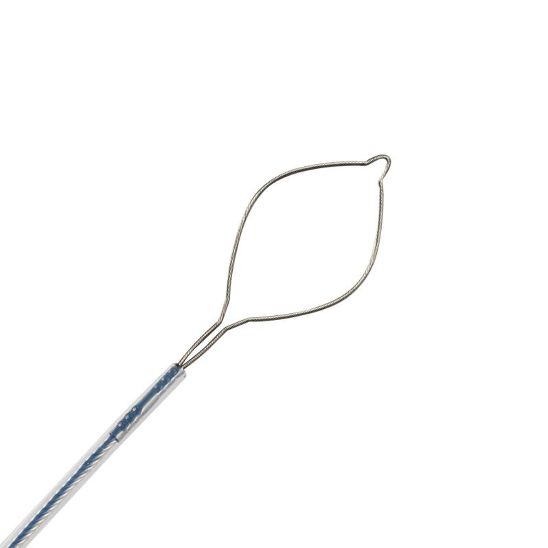
Disposable Polypectomy Snare
Examples of EMR
a. White light image
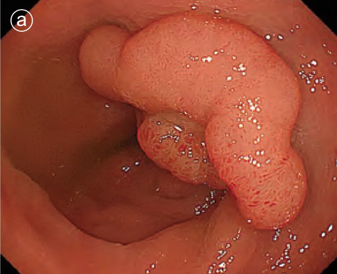
A 25 mm type IIa lesion with a slightly depressed central portion.
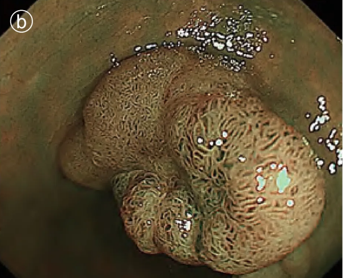
b. Narrow band imaging (NBI) images
c. Spraying indigo carmine to enlarge the image
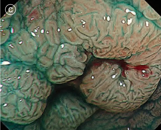
It was discovered that the depressions that were perceived by conventional observation were actually grooves between the leaves.
d. Enlarged image of crystal violet staining
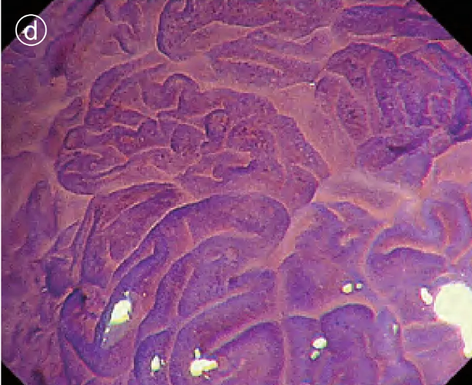
The pit pattern of the glandular duct opening at the edge of the lesion was type IV.
e. Enlarged image of crystal violet staining
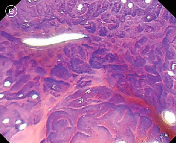
in the center of the lesion was VI, slightly irregular, and no obvious submucosal infiltration was found .
f. Local injection
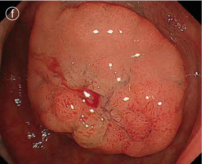
Puncture and local injection were performed in the center of the lesion , resulting in a good bulge.
g. Open the snare
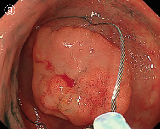
Press the snare tip against the colon wall to open the snare.
h. Close the snare
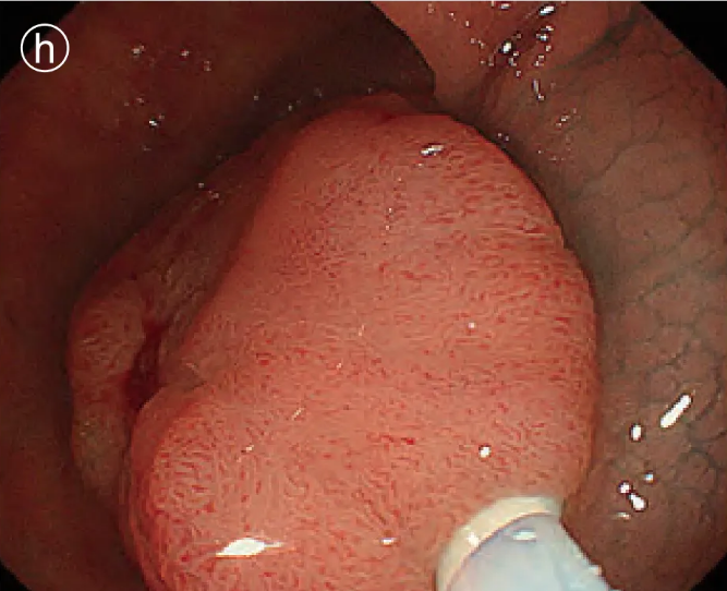
Close the snare and grasp the lesion.
i.Power on removal
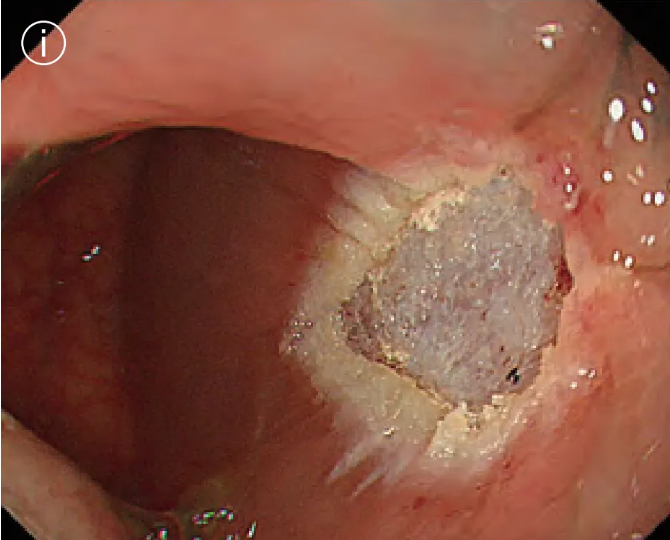
No perforation, bleeding or residual tumor was found.
j. Specimen fixation
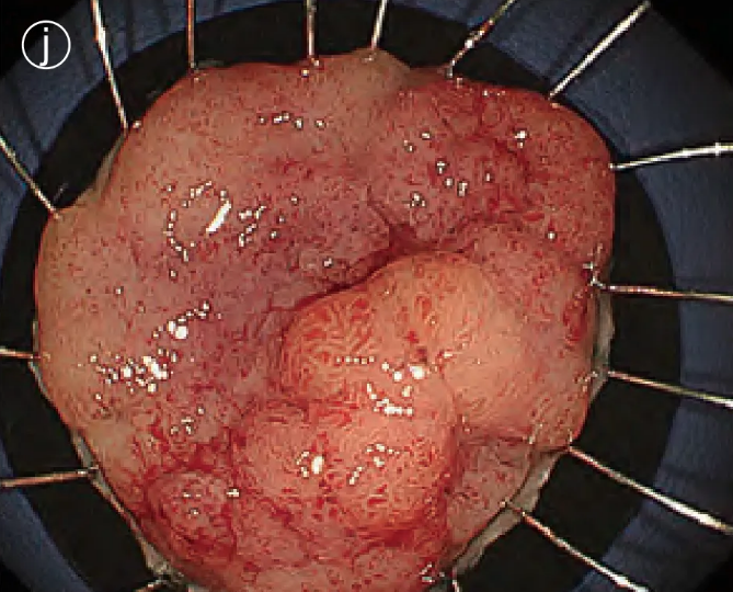
The excised specimen was attached to a rubber sheet.
Final pathological diagnosis: intramucosal carcinoma (Tis)
4.Tips for snare operation
The snare tip is gently placed on the oral mucosa of the lesion, then slowly opened and the snare root is pressed against the anal side of the lesion. To prevent the lateral incision from being positive, a small amount of normal mucosa should be inserted.
It should be noted that when the snare tip cannot be seen, it is possible that more normal mucosa has been inserted than expected. After the snare is fully tightened, push and pull the outer sleeve of the snare to observe the mobility of the lesion. If it is inserted into the muscular layer, the mobility of the lesion will be reduced.
Tips for electroresection
Do not press the snare against the bowel wall, but slightly elevate the lesion for resection. The risk of delayed perforation is low when using electrosurgical resection , but it is prone to intraoperative (early after resection) bleeding.
Excision that is too fast may cause bleeding, while excision that is too slow may cause delayed perforation. If the patient feels pain, or the assistant feels that the tissue is elastic like rubber and difficult to cut, it is likely that the tissue is involved in the muscle layer, and the excision should be stopped immediately.
Essentials for Endoscopy Technicians
If the endoscopist feels that the tissue is elastic like rubber and difficult to cut, he or she should immediately inform the operator to discuss countermeasures.
Tips for Sharding EMR
For larger lesions, it is sometimes safer to perform a piecemeal resection rather than a forced all-at-once resection. However, the more pieces there are, the greater the likelihood of local residual recurrence. Even with piecemeal EMR, initial resections should be performed as large as possible with a large snare to minimize the number of pieces.
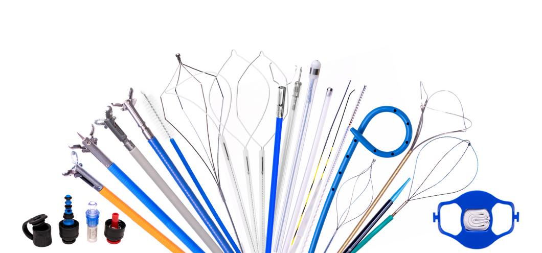
We, Jiangxi Zhuoruihua Medical Instrument Co.,Ltd., is a manufacturer in China specializing in the endoscopic consumables, such as biopsy forceps, hemoclip, polyp snare, sclerotherapy needle, spray catheter, cytology brushes, guidewire, stone retrieval basket, nasal biliary drainage catheter etc. which are widely used in EMR, ESD, ERCP. Our products are CE certified, and our plants are ISO certified. Our goods have been exported to Europe, North America, Middle East and part of Asia, and widely obtains the customer of the recognition and praise!
Biopsy forceps:
https://www.zrhendoscopy.com/single-use-endoscopic-tissue-biopsy-forceps-with-graduation-product/
Hemoclip
https://www.zrhendoscopy.com/disposable-rotatable-endoscopic-hemoclip-for-gastroscopy-use-product/
polyp snare
https://www.zrhendoscopy.com/disposable-endoscopic-resection-polypectomy-snare-for-gastroenterology-product/
sclerotherapy needle
https://www.zrhendoscopy.com/gastroenterology-accessories-endoscopic-sclerotherapy-injection-needle-product/
Spray catheter
https://www.zrhendoscopy.com/ce-certified-disposable-endoscopic-spray-catheter-for-digestive-chromoendoscopy-product/
cytology brushes
https://www.zrhendoscopy.com/endoscopy-accessories-disposable-endoscopic-cytology-brush-for-gastrointestinal-tract-product/
stone retrieval basket
https://www.zrhendoscopy.com/ercp-instrument-gallstone-stone-retrieval-basket-for-endoscopy-product/
nasal biliary drainage catheter
https://www.zrhendoscopy.com/medical-instrument-disposable-nasal-biliary-drainage-catheter-for-ercp-operation-product/
EMR
https://www.zrhendoscopy.com/emresd/
ESD
https://www.zrhendoscopy.com/emresd/
ERCP
https://www.zrhendoscopy.com/ercp/
Post time: Feb-13-2025